

1 Departamento de Medicamentos-Faculdade de Farmácia, Universidade Federal do Rio de Janeiro – UFRJ, Rio de Janeiro, Brazil
2 Instituto de Microbiologia Professor Paulo de Góes, Universidade Federal do Rio de Janeiro – UFRJ, Rio de Janeiro, Brazil
3 Instituto de Ciências Biomédicas, Universidade Federal do Rio de Janeiro – UFRJ, Rio de, Janeiro, Brazil
4 Departamento de Fármacos-Faculdade de Farmácia, Universidade Federal do Rio de Janeiro – UFRJ, Rio de Janeiro, Brazil
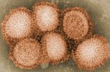
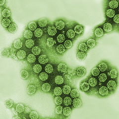
 O bioterápico denominado Influenzinum RC, foi preparado através da ultradiluição (diluição homeopática 30dH) do vírus Influenza A (A/Aichi/2/68 H3N20). As alterações celulares induzidas por este bioterápico foram analisadas através de analises bioquímicas e microscopia óptica e eletrônica.
O Influenzinum RC não causou efeito citotóxico nem alterações morfológicas em célula MDCK (Madin–Darby canine kidney) Apos 30 dias foi observado um aumento na taxa de mitose. A atividade mitocondrial foi alterada após tratamento com 10 e 30 dias. Ocorreu diminuição de PFK-1 e em células de macrófagos J774.48 tratadas a citocina TNF-α aumentou nos sobrenadantes das culturas de macrófagos.
H3N2 homeopathic influenza virus solution modifies cellular and biochemical aspects of MDCK and J774G8 cell lines
Objectives
To develop a biotherapy prepared from the infectious influenza A virus (A/Aichi/2/68 H3N2) and to verify its in vitro response.
Methods
The ultradiluted influenza virus solution was prepared in the homeopathic dilution 30dH, it was termed Influenzinum RC. The cellular alterations induced by this preparation were analyzed by optical and electron microscopy, MTT and neutral red assays. Glycolytic metabolism (PFK-1) was studied by spectrophotometric assay. Additionally, the production of tumor necrosis factor-α (TNF-α) by J774.G8 macrophage cells was quantified by ELISA before and after infection with H3N2 influenza virus and treatment
Results
Influenzinum RC did not cause cytotoxic effects but induced morphological alterations in Madin–Darby canine kidney (MDCK) cells. After 30 days, a significant increase (p < 0.05) in mitosis rate was detected compared to control. MDCK mitochondrial activity was changed after treatment for 10 and 30 days. Treatment significantly diminished (p < 0.05) PFK-1 activity. TNF-α in biotherapy-stimulated J774.G8 macrophages indicated a significant (p < 0.05) increase in this cytokine when the cell supernatant was analyzed.
Conclusion
Referencia: Homeopathy 2013; 102(1);31-40
 Por Tatiana F. Robaina,Gabriella S. Mendes,Fabrıcio J. Benati,Giselle A. Pena, Raquel C. Silva, Miguel A.R. Montes,Maria Elisa R. Janini,Fernando P. Câmara e Norma Santos.
Este estudo é o resultado de uma colaboração entre o Instituto de Microbiologia e a escola de Odontologia, Universidade Federal do Rio de Janeiro. Através de analises por PCR foi detectada a presença de polyomavirus na saliva de indivíduos saudáveis. Das 291 amostras testadas 71(24.3%) foram positivas pra pelo menos um dos polyomavirus testados.
A detecção foi mais alta, principalmente, em indivíduos com 15-19 anos de idade (46.0%; 23/50) e em uma escala menor em pessoas com 50 anos de idade (33.3%; 9/27). Estes resultados reforçam a hipótese de que a saliva pode ser uma via para a transmissão do BKV e que a cavidade oral pode ser um sitio de replicação viral. Outros polyomavirus podem ser transmitidos de modo similar.
Shedding of Polyomavirus in the Saliva of Immunocompetent Individuals
The aim of this study was to investigate and compare the frequency of BKV, JCV, WUV, andKIV in the saliva of healthy individuals. Samples were analyzed for the presence of polyomaviruses (BKV, JCV, WUV, and KIV) DNA byreal-time PCR. Of the 291 samples tested, 71 (24.3%) were positive for at least one of the screened polyomaviruses. Specifically, 12.7% (37/291) were positive for WUV, 7.2% (21/291) positive for BKV, 2.4% (7/291) positive for KIV, and 0.3% (1/291) positive for JCV. BKV and WUV co-infections were detected in 1.7% (5/291) of individuals. No other co-infection combinations were found. The mean number of DNA copies was high, particularly for WUV and BKV, indicating active replication of these viruses. Polyomavirus detection was higher among individuals 15–19 years of age (46.0%; 23/50) and 50 years of age (33.3%; 9/27). However, the detection rate in the first group was almost 1.7 % greater than the latter. WUV infections were more frequent in individuals between the ages of 15 and 19 years and the incidence decreased with age. By contrast, BKV excretion peaked and persisted during the third decade of life and KIV infections were detected more commonly in subjects 50 years old. These findings reinforced the previous hypotheses that saliva may be a route for BKV transmission, and that the oral cavity could be a site of virus replication. These data also demonstrated that JCV, WUV, and KIV may be transmitted in a similar fashion.
J. Med. Virol. 85:144–148,2013. http://onlinelibrary.wiley.com/doi/10.1002/jmv.23453/pdf
 Por Fernando Portela Câmaraa, Luiz Max de Carvalhoa, and Ana Luisa Bacellar Gomesb
aSector for Infectious Diseases Epidemiology, Institute of Microbiology, Federal University of Rio de Janeiro (UFRJ), Health Sciences Center - Block I, University City - Fundão Island, Rio de Janeiro - RJ - CEP: 21941-590, Brazil
bInstituto de Medicina Social, Universidade do Estado do Rio de Janeiro (UERJ), Rio de Janeiro, Brazil
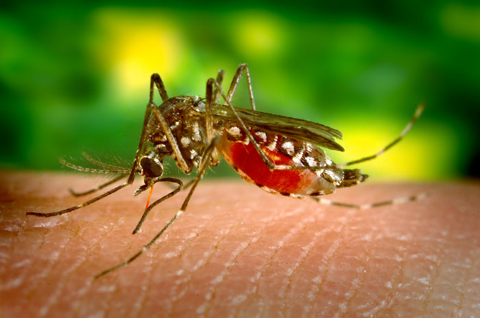
A febre amarela é uma arbovirose que no Brasil ocorre apenas na forma silvestre, Neste trabalho foi realizada uma analise demográfica de 831 casos que ocorreram no Brasil no período de 1973-2008 para determinar os grupos de risco da população. O estudo identificou homens adultos, com 30 anos, moradores de áreas rurais nos estados do Pará, Goiás, Maranhão e Minas Gerais que não foram vacinados ou cuja vacina estava vencida como o principal grupo doença. Dos casos notificados ocorreu uma taxa de mortalidade de 51% ( 421/831).
Demographic profile of sylvatic yellow fever (SYF) in Brazil from 1973 to 2008
Yellow fever is an acute, frequently fatal, febrile arbovirosis that in Brazil occurs only in the sylvatic form. Sylvatic yellow fever (SYF) appears in sporadic outbreaks over a large area of Brazil. In this paper, we analyze the demographic profile of 831 SYF cases that occurred between 1973 and 2008, to determine which segments of the exposed population are at greater risk.
Trans R Soc Trop Med Hyg (2013)doi: 10.1093/trstmh/trt014
In press http://trstmh.oxfordjournals.org/content/early/2013/02/25/trstmh.trt014.abstract
